V Fib Ecg Strip
Ventricular Fibrillation Vf Litfl Ecg Library Diagnosis
Ventricular Fibrillation Acls Algorithms Com
Ventricular Fibrillation Vf Litfl Ecg Library Diagnosis

Acls Ventricular Fibrillation And Pulseless Ventricular Tachycardia Guide
Ventricular Fibrillation Acls Algorithms Com

Ventricular Fibrillation Wikipedia

Ventricular fibrillation is an emergency condition requiring immediate action.
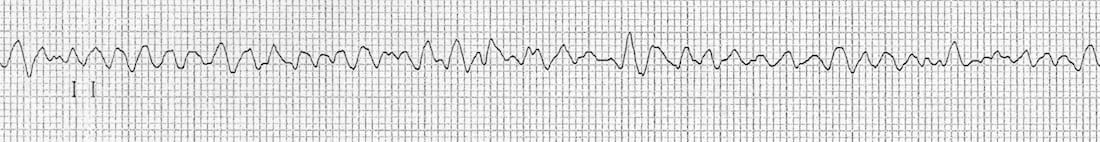
V fib ecg strip. These features include observing p wave forms measurement of ekg intervals and segments assessment of rhythm calculating heart rate and the evaluation of other relevant wave segments. By bagnall et al. Ventricular fibrillation is always pulseless and must be confirmed by ekg or defibrillator monitor. Symptoms of both afib and vfib are shortness of breath dizziness nausea and chest pain.
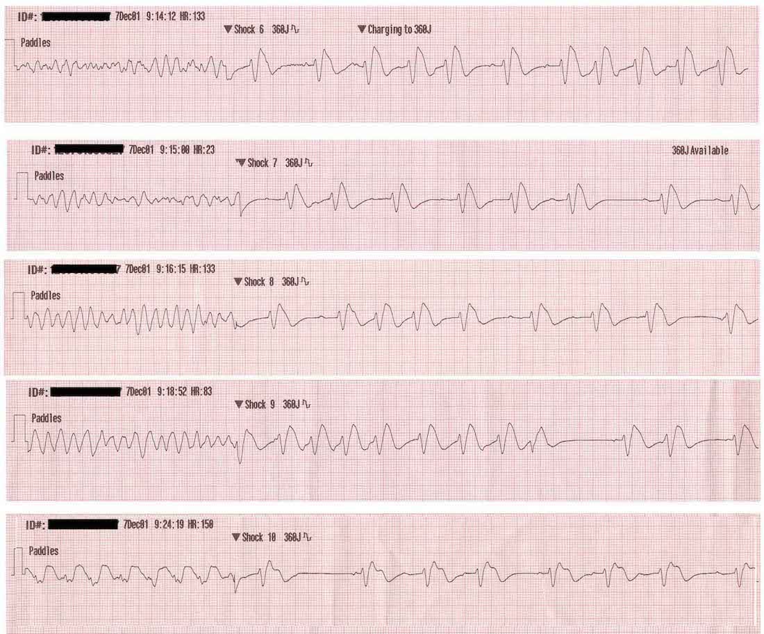
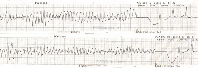
This is followed by death in the absence of treatment. The course provides training on the key features of an ekg tracing. When the ventricles handle the pacemaking role they can be observed on ekg tracings. Defibrillation is the treatment of choice and should occur as soon as possible.
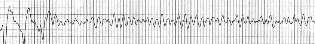
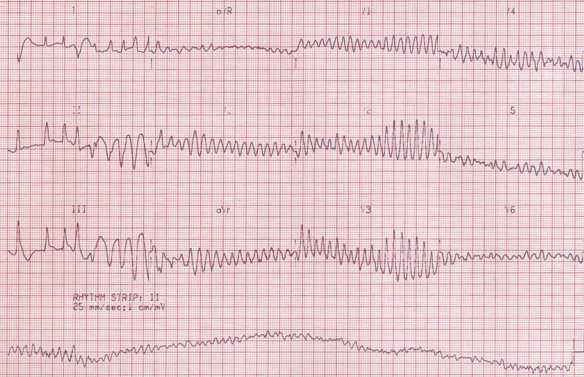
Ecg showing ventricular tachycardia degenerating into ventricular fibrillation. 12 lead and rhythm strip. The rate will be 120 200 bpm. A good starting point for learning about v fib and other types of ekg interpretation is our ekg basics training course.
Ventricular fibrillation is treated according to the resuscitation algorithm. Cardiac arrest and sudden cardiac death. Atrial fibrillation afib and ventricular fibrillation vfib are both a type of abnormal heart rhythm arrhythmia. Second degree av block type i.
Av block and st elevation. Ventricular fibrillation is initially found in about 10 of. Atrial fibrillation is caused by irregular electrical impulses in the atria and ventricular fibrillation is caused by irregular electrical impulses in the ventricles. The mother rotor then gives rise to propagating unstable daughter wavefronts which results in the chaotic electrical activity seen on the ecg.
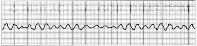
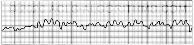
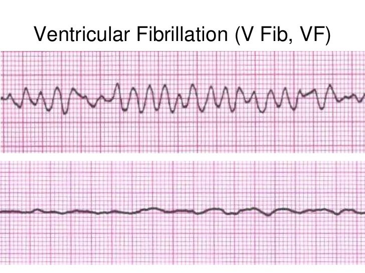
The fibrillation is maintained by re entry circuits formed by some of the wavelets. Ventricular fibrillation v fib or vf is an abnormal heart rhythm in which the ventricles of the heart quiver instead of pumping normally. The ecg criteria to diagnose ventricular fibrillation vfib on a 12 lead ecg is discussed including a brief discussion of treatment using automated external defibrillators aed and icds. The video below shows an example of what ventricular fibrillation will look like when you see it on the defibrillator monitor.
Ventricular tachycardia a sequence of three pvcs in a row is ventricular tachycardia. It is due to disorganized electrical activity. Mother rotor mechanism in which a stable re entry circuit is formed the mother rotor.
Ventricular Fibrillation Vf Litfl Ecg Library Diagnosis

Acls Ventricular Fibrillation And Pulseless Ventricular Tachycardia Guide

Dr Smith S Ecg Blog Ventricular Fibrillation On A 12 Lead Ecg
Ventricular Tachycardia Vt Ecg Review Criteria And Examples Learntheheart Com

V Fib Ecg Guru Instructor Resources

Matters Of The Heart Cardiac Arrest Patmac Rn

From Atrial Fibrillation To Ventricular Fibrillation And Back Circulation
Ventricular Fibrillation Vf Litfl Ecg Library Diagnosis
Basic Ekg And Rhythm Interpretation Symposia The Crudem Foundation
Http Keymedinfo Com Site 667keym Cardiac Dysrhythmia Overview To Help With Acls Precourse Examination Pdf

Atrial Fibrillation Vs Ventricular Fibrillation What S The Difference
V Tach Ecg Guru Instructor Resources
Http Keymedinfo Com Site 667keym Cardiac Dysrhythmia Overview To Help With Acls Precourse Examination Pdf
